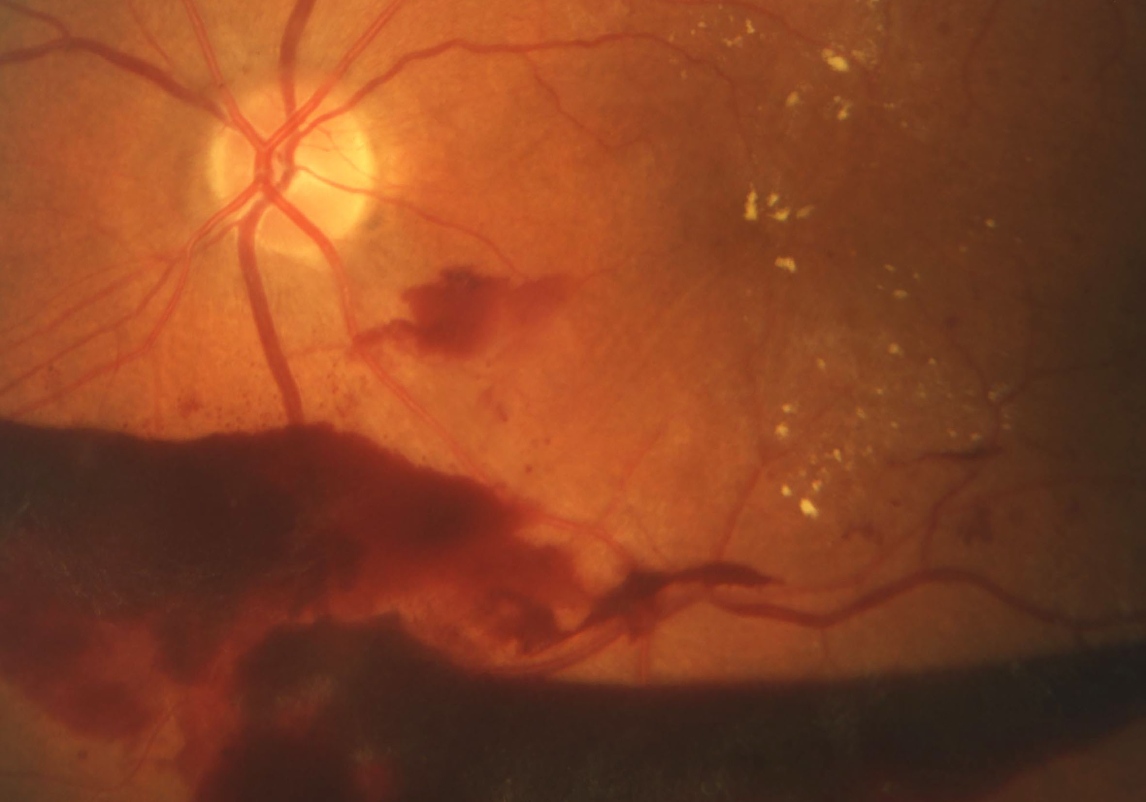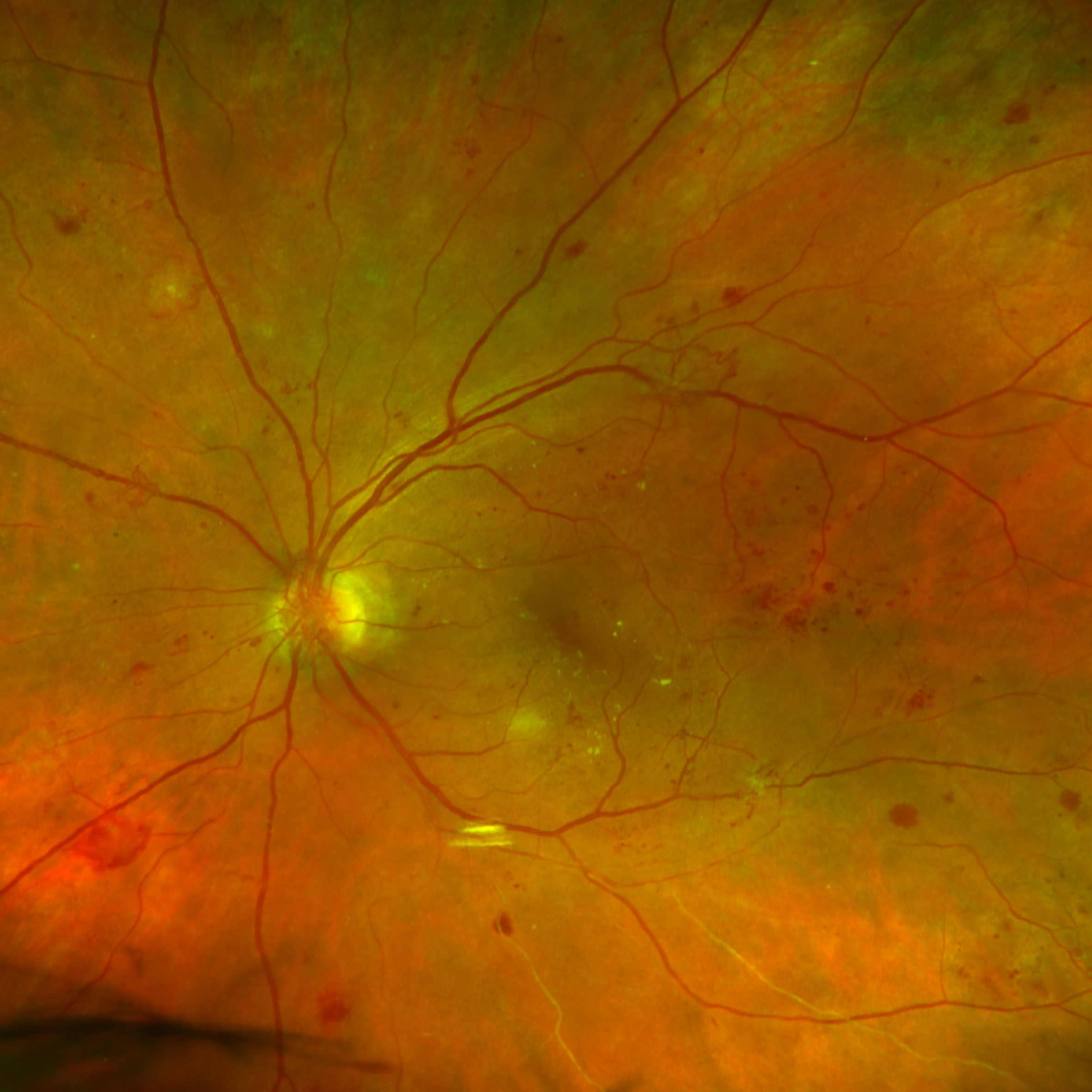
Milde diabetische Augenbeteiligung mit Blutungen in der Netzhaut
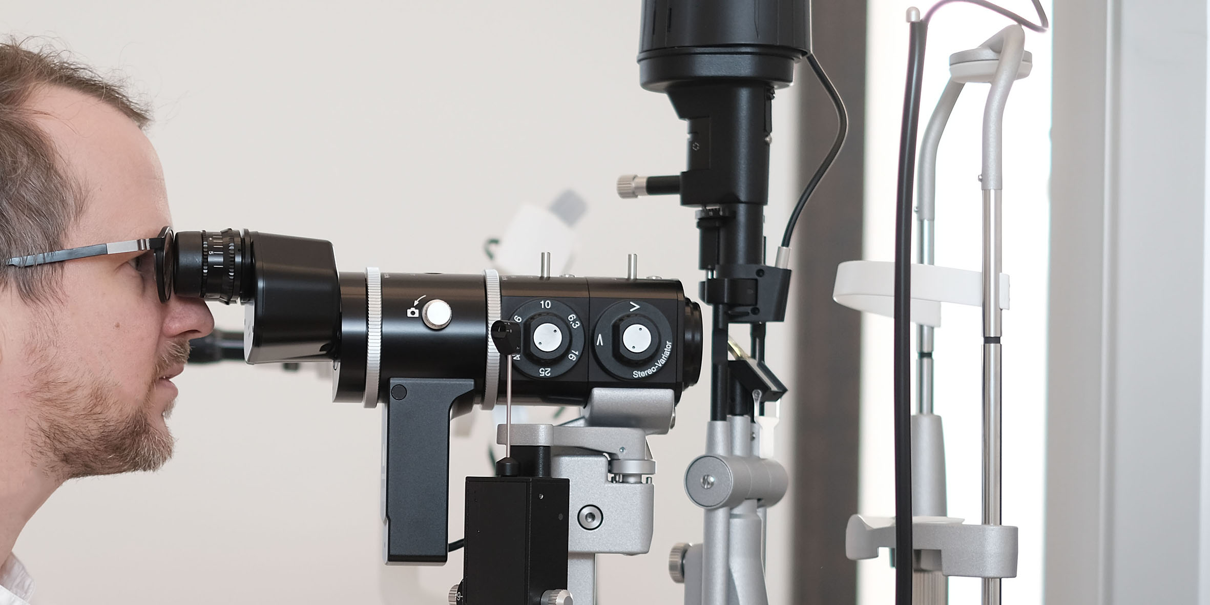
Although people suffering from diabetes are at higher risk of getting blind, the majority of them usually have none or only mild ocular involvement. Regular eye examination is important for early detection of eye deterioration. If a serious eye condition occurs, there are several forms of therapy that show a high success rate.
What kind of eye damage can occur in diabetes?
Glaucoma, cataract and retinopathy are the eye diseases that can occur in diabetes.
Glaukom – Grüner Star
Diabetics have a higher risk of developing a cataract. The longer someone has diabetes, the greater the risk of developing glaucoma. Therefore, the pressure in the eye and the optic nerve should be monitored, and if necessary visual field tests should be done regularly.
Cataract
Patients suffering from diabetes may develop cataracts earlier and cataract can progress faster. The operation is performed as in non-diabetics.
Diabetic retinopathy – retinal disease
This is a general term for all diseases that can occur on the retina in patients with diabetes. The following diseases are summarized under this term:
Nonproliferative retinopathy
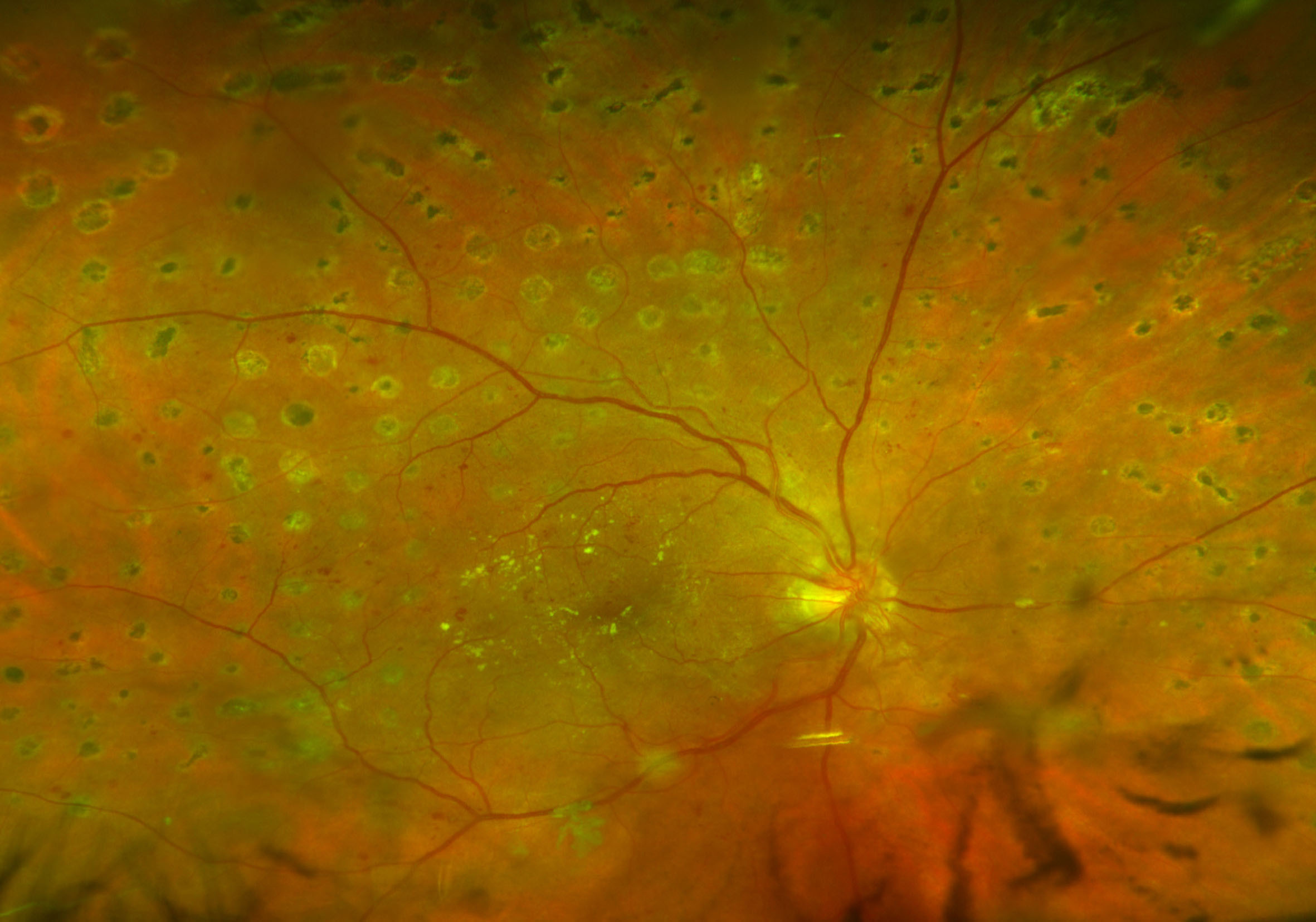
It is the most common form of retinopathy. This changes the smallest vessels in the retina (capillaries), which form extensions and enlarge in a balloon form. The proliferative retinopathy is divided into three stages depending on how many blood vessels are no longer consistent and how much of the retina is no longer supplied with oxygen and nutrients.
Diabetic macular edema
The small blood vessels in the eye can be damaged by the diabetes so that the fluid exchange between vessels and retina does not work properly. Liquid can therefore accumulate more and more in the area of the sharpest vision, the macula, and lead to a loss of vision. This is called diabetic Makulödem.
Proliferative retinopathy
This is the most pronounced form of diabetic retinopathy, which can develop in the course of several years. In this disease, the blood vessels found in the eye are damaged in such an extent that they are partially completely closed. In response to this, new but inferior vessels form in the retina. These can cause bleeding, a vitreous hemorrhage (vitreous = transparent gel inside the eye) and the patient’s vision is more severely restricted. The inferior vessels may also favor the development of scar tissue in the eye. If this subsequently begins to shrink, a retinal detachment (tractive retinal detachment) may occur.
Therapy
- Laser coagulation of the retina
- Intravitreal injections
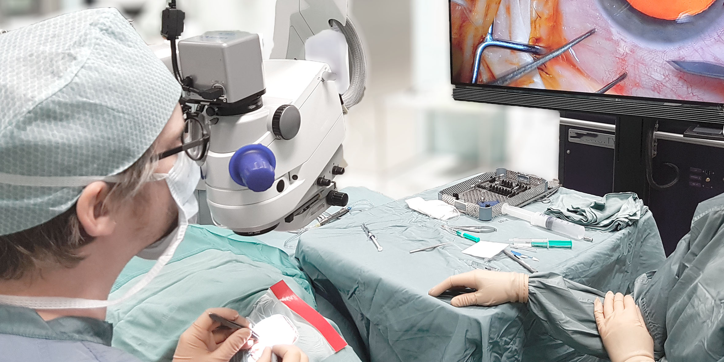
- Vitrectomy


